View the images and select the correct diagnosis from the list below.
You answered: Endometrial cells
Sorry, that is incorrect.
The correct diagnosis is: Small cell carcinoma
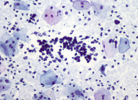
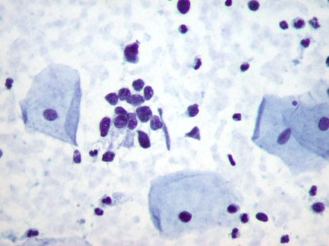
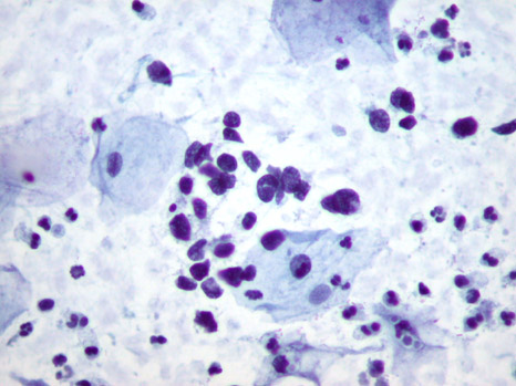
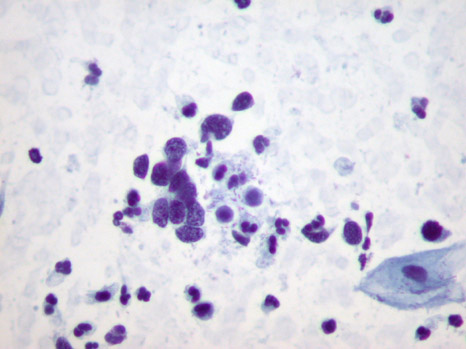
- The smear contains numerous groups of loosely cohesive aggregates of small cells with scant cytoplasm.
- The nuclei are hyperchromatic and angular. Nuclear moulding is evident.
- The cytologic features suggest small cell carcinoma.
- Small cell carcinoma is a highly aggressive tumour of neuroendocrine differentiation strongly associated with HPV types 18 and 16.
- The routine cervical smear is a relatively insensitive and nonspecific method of detecting small cell carcinoma. The specific diagnosis on cervical smears is difficult. Small cell carcinoma can mimic follicular cervicitis, endometrial cells, endocervical adenocarcinoma, squamous cell carcinoma of small cell type, and non-Hodgkin's lymphoma. Suspicion should prompt an urgent biopsy to establish the diagnosis and initiate prompt treatment.
- Biopsies reveal tumours composed of densely packed small to intermediate sized cells with scant cytoplasm, oval to spindle shaped, hyperchromatic nuclei and usually indistinct nucleoli. Mitoses and apoptotic nuclear debris are frequent.
Back to images
- Bonfiglio, T.A. & Erozan, Y.S. (1997).
Gynecologic Cytopathology. Philadelphia: Lipincott-Raven 145.
- Zhou, C., Hayes, M. M., Clement, P. B. & Thomson, T. A. (1998). Small cell carcinoma of the uterine cervix: cytologic findings in 13 cases.
Cancer Cytopathology 85(5), 281-8.
You answered: Marked squamous dyskaryosis.
Sorry, that is
incorrect.
The correct diagnosis is: Small cell carcinoma
- The smear contains numerous groups of loosely cohesive aggregates of small cells with scant cytoplasm.
- The nuclei are hyperchromatic and angular. Nuclear moulding is evident.
- The cytologic features suggest small cell carcinoma.
- Small cell carcinoma is a highly aggressive tumour of neuroendocrine differentiation strongly associated with HPV types 18 and 16.
- The routine cervical smear is a relatively insensitive and nonspecific method of detecting small cell carcinoma. The specific diagnosis on cervical smears is difficult. Small cell carcinoma can mimic follicular cervicitis, endometrial cells, endocervical adenocarcinoma, squamous cell carcinoma of small cell type, and non-Hodgkin's lymphoma. Suspicion should prompt an urgent biopsy to establish the diagnosis and initiate prompt treatment
- Biopsies reveal tumours composed of densely packed small to intermediate sized cells with scant cytoplasm, oval to spindle shaped, hyperchromatic nuclei and usually indistinct nucleoli. Mitoses and apoptotic nuclear debris are frequent.
Back to images
- Bonfiglio, T.A. & Erozan, Y.S. (1997).
Gynecologic Cytopathology. Philadelphia: Lipincott-Raven 145.
- Zhou, C., Hayes, M. M., Clement, P. B. & Thomson, T. A. (1998). Small cell carcinoma of the uterine cervix: cytologic findings in 13 cases.
Cancer Cytopathology 85(5), 281-8.
You answered: Small cell carcinoma.
That is correct!!
- The smear contains numerous groups of loosely cohesive aggregates of small cells with scant cytoplasm.
- The nuclei are hyperchromatic and angular. Nuclear moulding is evident.
- The cytologic features suggest small cell carcinoma.
- Small cell carcinoma is a highly aggressive tumour of neuroendocrine differentiation strongly associated with HPV types 18 and 16.
- The routine cervical smear is a relatively insensitive and nonspecific method of detecting small cell carcinoma. The specific diagnosis on cervical smears is difficult. Small cell carcinoma can mimic follicular cervicitis, endometrial cells, endocervical adenocarcinoma, squamous cell carcinoma of small cell type, and non-Hodgkin's lymphoma. Suspicion should prompt an urgent biopsy to establish the diagnosis and initiate prompt treatment
- Biopsies reveal tumours composed of densely packed small to intermediate sized cells with scant cytoplasm, oval to spindle shaped, hyperchromatic nuclei and usually indistinct nucleoli. Mitoses and apoptotic nuclear debris are frequent.
Back to images
- Bonfiglio, T.A. & Erozan, Y.S. (1997).
Gynecologic Cytopathology. Philadelphia: Lipincott-Raven 145.
- Zhou, C., Hayes, M. M., Clement, P. B. & Thomson, T. A. (1998). Small cell carcinoma of the uterine cervix: cytologic findings in 13 cases.
Cancer Cytopathology 85(5), 281-8.
You answered: Endocervical adenocarcinoma.
Sorry, that is incorrect.
The correct diagnosis is: Small cell carcinoma.
- The smear contains numerous groups of loosely cohesive aggregates of small cells with scant cytoplasm.
- The nuclei are hyperchromatic and angular. Nuclear moulding is evident.
- The cytologic features suggest small cell carcinoma.
- Small cell carcinoma is a highly aggressive tumour of neuroendocrine differentiation strongly associated with HPV types 18 and 16.
- The routine cervical smear is a relatively insensitive and nonspecific method of detecting small cell carcinoma. The specific diagnosis on cervical smears is difficult. Small cell carcinoma can mimic follicular cervicitis, endometrial cells, endocervical adenocarcinoma, squamous cell carcinoma of small cell type, and non-Hodgkin's lymphoma. Suspicion should prompt an urgent biopsy to establish the diagnosis and initiate prompt treatment
- Biopsies reveal tumours composed of densely packed small to intermediate sized cells with scant cytoplasm, oval to spindle shaped, hyperchromatic nuclei and usually indistinct nucleoli. Mitoses and apoptotic nuclear debris are frequent.
Back to images
- Bonfiglio, T.A. & Erozan, Y.S. (1997).
Gynecologic Cytopathology. Philadelphia: Lipincott-Raven 145.
- Zhou, C., Hayes, M. M., Clement, P. B. & Thomson, T. A. (1998). Small cell carcinoma of the uterine cervix: cytologic findings in 13 cases.
Cancer Cytopathology 85(5), 281-8.
From the cytopathology files of BC Cancer.
Submitted by: Brenda Smith, BSc and Tom Thomson, MD.


